 Founded in 1980 |
|
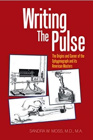
| Sandra W. Moss, MD, MA, Writing the Pulse: The Origins and Career of the Sphygmograph and Its American Masters, XLibris, 2018. ISBN 978-1-5434-6358-3. |
Reviewer: Allen B. Weisse, MD, FACC
June 6, 2018
As far back as antiquity physicians have studied the pulse as an indicator of disease.
Portraiture of the Middle Ages and the Renaissance represented physicians with their "educated" fingers palpating the wrists of their patients, their intense concentration on the pulse registered in their features. It was not until the mid-nineteenth century, however, that physicians began to explore some form of recording to report their findings objectively.
What made this possible was the invention of the kymograph by Carl Ludwig, at the University of Leipzig, in the 1840's. This instrument, with its revolving drum, could record physiological events over a short time with inscriptions being made by a stylus scratching out a pattern over paper previously blackened with smoke. The sphygmograph, utilizing this technology, obtained tracings that reflected the changing size of the radial artery during the course of cardiac contraction and relaxation.
Medical historian Sandra W. Moss is not a cardiologist—she is board-certified in nephrology—but she has, nonetheless, provided us with the most comprehensive work on this subject that we are likely to find now or in the future. The book contains more than 350 bibliographic sources, reflects the contributions of more than two dozen individuals involved in the story, and includes 65 illustrations depicting various models that were introduced over time. Lacking, however, are photographs of the pioneers, which might have added a little more "skin and bones" to the enterprise.
The construction and the application of the sphygmograph were relatively simple. A sensor was placed over the radial artery, partially compressing it, to find the desired wave pattern, one similar to those seen today on monitors in the operating suite or intensive care unit. Despite the relative ease with which these recordings were made, the patterns could be altered by injudicious application of pressure over the artery, movement of the patient’s arm, and other factors. Criteria determined by one physician may not have been the same as for others. Most importantly, there was no universal unit of measurement, the tracing simply reflecting the changing of arterial diameter over the cardiac cycle. Finally, some skill and much study were required from the practitioner in mastering the technique.
Use of ink pens and rolls of paper allowed for longer intervals of operation. Later on, the "polygraph" was introduced, with which multiple recordings could be made simultaneously, including venous waves, the apical impulse, and the electrocardiogram.
By the beginning of the twentieth century, blood pressure values could be obtained, thanks to the work of Riva-Rocci and Koratkoff, their sphygmomanometers determining pressure in millimeters of mercury. X-rays of the heart, angiography, cardiac catheterization, and echo Doppler recordings sealed the fate of the sphygmograph. Moss skillfully introduces such considerations toward the end of her narrative. Yet, she points out, despite its limitations, sphygmography had an undeniable role in the demand for and development of graphic techniques in clinical medicine.
James Mackensie, probably one of the most successful of those using the technique, cautioned against overreliance upon it. Clinical acumen was still required. Today, with sphygmography long gone, we continue to rely on findings so well documented by the technique. Despite the dominance of the technological revolution, our "educated" fingers can still detect pulsus paradoxus in cardiac tamponade; pulsus parvus et tardus in aortic stenosis; pulsus alternans in cardiac failure; the collapsing water hammer pulse of aortic regurgitation; the double-humped bisferiens pulse of hypertrophic subaortic stenosis; and the irregularly irregular pulse of atrial fibrillation. All awaiting us at the bedside.
